Comprehensive Heart Ultrasound
At Phoenix Ultrasound, we provide advanced echocardiography, a safe, non-invasive ultrasound scan that evaluates the structure and function of your heart. This test uses high-frequency sound waves to produce detailed images of your heart chambers, valves, and blood flow, helping your doctor assess how well your heart is working.
Hear the summary of the text below from the following voice:
📋 What is an Echocardiogram?
An echocardiogram (echo), or heart ultrasound, is a diagnostic test that uses sound waves to create live moving images of the heart.
It helps detect abnormalities in heart size, shape, movement, and function. The test is completely painless and uses no radiation
| Feature | Details |
|---|---|
| Test | Echocardiogram / Heart Ultrasound |
| Purpose | Assess heart structure, function, valves, and blood flow |
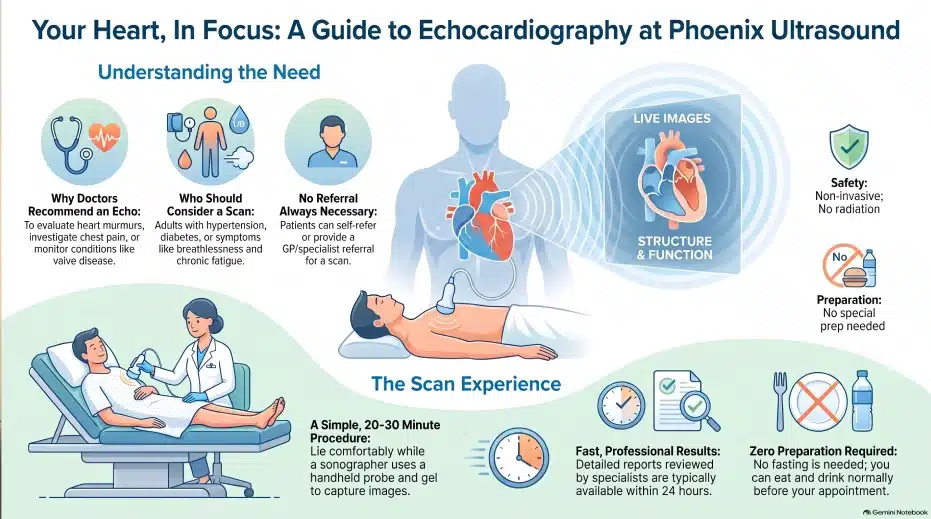
| Duration | Approximately 20–30 minutes |
| Radiation | None |
| Invasiveness | Non-invasive and painless |
| Preparation | No special preparation required; eat and drink normally |
| Referral | Self-referral or GP/specialist referral |
| Results | Detailed report usually available within 24 hours |
| Who It’s For | People with heart symptoms, cardiovascular risk factors, or those needing heart health assessment |
Why is Echocardiography Done?
Your doctor may recommend an echocardiogram to:
- ✔ Evaluate heart murmurs or abnormal sounds
- ✔ Investigate chest pain, shortness of breath, or palpitations
- ✔ Assess damage after a heart attack
- ✔ Monitor known heart conditions such as valve disease, cardiomyopathy, or heart failure
- ✔ Check heart function before or after major surgery
- ✔ Screen for inherited cardiac disorders
Who Should Consider an Echocardiogram?
This scan is suitable for:
- ✔ Adults with cardiovascular risk factors (e.g. hypertension, diabetes, family history of heart disease)
- ✔ Patients with symptoms like breathlessness, chest pain, or fatigue
- ✔ Individuals with previous abnormal ECG or chest X-ray findings
- ✔ Patients on long-term chemotherapy or medications affecting the heart
- ✔ Those seeking a routine heart health assessment or preventive screening
What Happens During the Scan?
This scan is suitable for:
- ✔ The procedure takes about 20-30 minutes.
- ✔ You will lie comfortably on an examination couch.
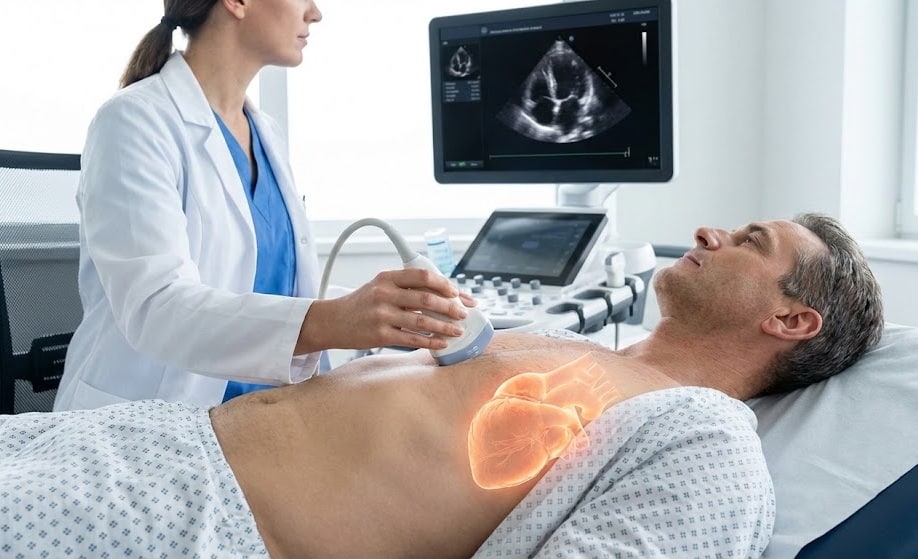
- ✔ A small amount of gel will be applied to your chest, and the sonographer will move a handheld probe over the area to capture heart images.
- ✔ You may be asked to change positions or briefly hold your breath to obtain clearer images.
- ✔ After the scan, the results are reviewed by a specialist cardiac sonographer and consultant cardiologist if required.
Key Benefits
- ✅ Non-invasive and completely safe (no radiation)
- ✅ Real-time assessment of heart structure and blood flow
- ✅ Early detection of heart disease
- ✅ Fast and accurate results
- ✅ Professional reporting by experienced cardiac sonographers
Key Points to Remember
- ✔ Preparation: No special preparation is needed. You can eat and drink as normal.
- ✔ Duration: Approximately 20-30 minutes.
- ✔ Results: A detailed report is usually available within 24 hours.
- ✔ Referral: You can self-refer or come with a GP/specialist referral.
📅 Book Your Scan Today
📞 Call us: 020 3318 1373
📧 Email: Contact@phoenix-ultrasound.co.uk
📍 Our Locations:
Central London: 1-5 Portpool Lane, London, Medical Room, EC1N 7UU
Surrey: 63 Nork Way, Banstead, SM7 1HL