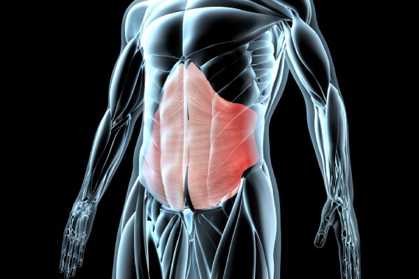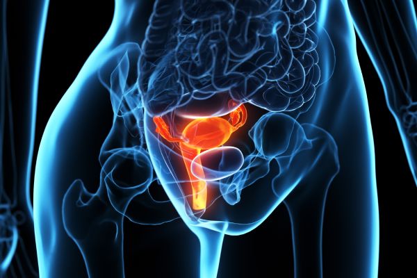
Prostate Transrectal Shear Wave Elastography & Colour Doppler(Multi Parametric)

Price : £465
Book an appoinment
Prostate Transrectal Ultrasound Scan with Shear Wave Elastography & Colour Doppler (Multi-Parametric)
Prostate Shear Wave Elastography (SWE) is a cutting-edge, non-invasive imaging technique designed to assess the stiffness and vascular characteristics of the prostate. When combined with Colour Doppler, this multi-parametric approach significantly enhances the detection and characterization of prostate conditions — particularly prostate cancer, one of the most common cancers among men worldwide.
🧬 How It Works
Shear waves are generated within the prostate gland and their speed — which increases in stiffer (potentially cancerous) tissues — is measured and displayed as a colour-coded map (elastogram). Colour Doppler simultaneously evaluates blood flow, further improving diagnostic precision.
💡 Key Benefits
- ✔ Improved Diagnostic Accuracy: Differentiates between benign and malignant prostate lesions more reliably.
- ✔ Non-Invasive & Painless: A comfortable, patient-friendly alternative to invasive procedures.
- ✔ Reduces Unnecessary Biopsies: Helps identify high-risk areas before proceeding to invasive biopsy.
- ✔ Assists in Treatment Planning: Evaluates tumor aggressiveness to tailor therapy effectively.
🩺 Clinical Applications
- ✔ Early Screening: Ideal for men with elevated PSA or abnormal digital rectal exam (DRE) findings.
- ✔ Targeted Biopsy Planning: Guides needle placement to increase diagnostic yield.
- ✔ Active Surveillance: Monitors known prostate lesions over time for changes in stiffness or vascularity.
🕒 Procedure Overview
- ✔ Duration: Approximately 30–40 minutes
- ✔ No preparation required (unless otherwise advised)
- ✔ Results sent to your device and email
- ✔ Shareable with your urologist or GP
📅 Book Your Prostate SWE + Doppler Scan
If you’re experiencing urinary symptoms, elevated PSA, or simply prioritising proactive men’s health screening, this advanced ultrasound scan provides a thorough and accurate assessment of your prostate.
📞 Call us: 020 3318 1373
📧 Email: Contact@phoenix-ultrasound.co.uk
📍 Our Locations:
Central London: 1-5 Portpool Lane, London, Medical Room, EC1N 7UU
Surrey: 63 Nork Way, Banstead, SM7 1HL
Prostate Shear Wave Elastography is a sophisticated ultrasound imaging technique that measures the stiffness of prostate tissue. It generates shear waves within the prostate gland and analyzes their speed to determine tissue stiffness, which helps in identifying areas that may be affected by conditions such as prostate cancer.
While traditional prostate ultrasound relies on visual assessment of the prostate gland to identify abnormalities, Prostate SWE provides quantitative data on tissue stiffness. This additional information can improve the accuracy of distinguishing between benign and potentially malignant prostate lesions.
Prostate SWE may be recommended for men who have elevated PSA levels, abnormal findings on digital rectal exams, or those undergoing active surveillance for prostate cancer. It's particularly useful for identifying areas within the prostate that may require further evaluation through biopsy.
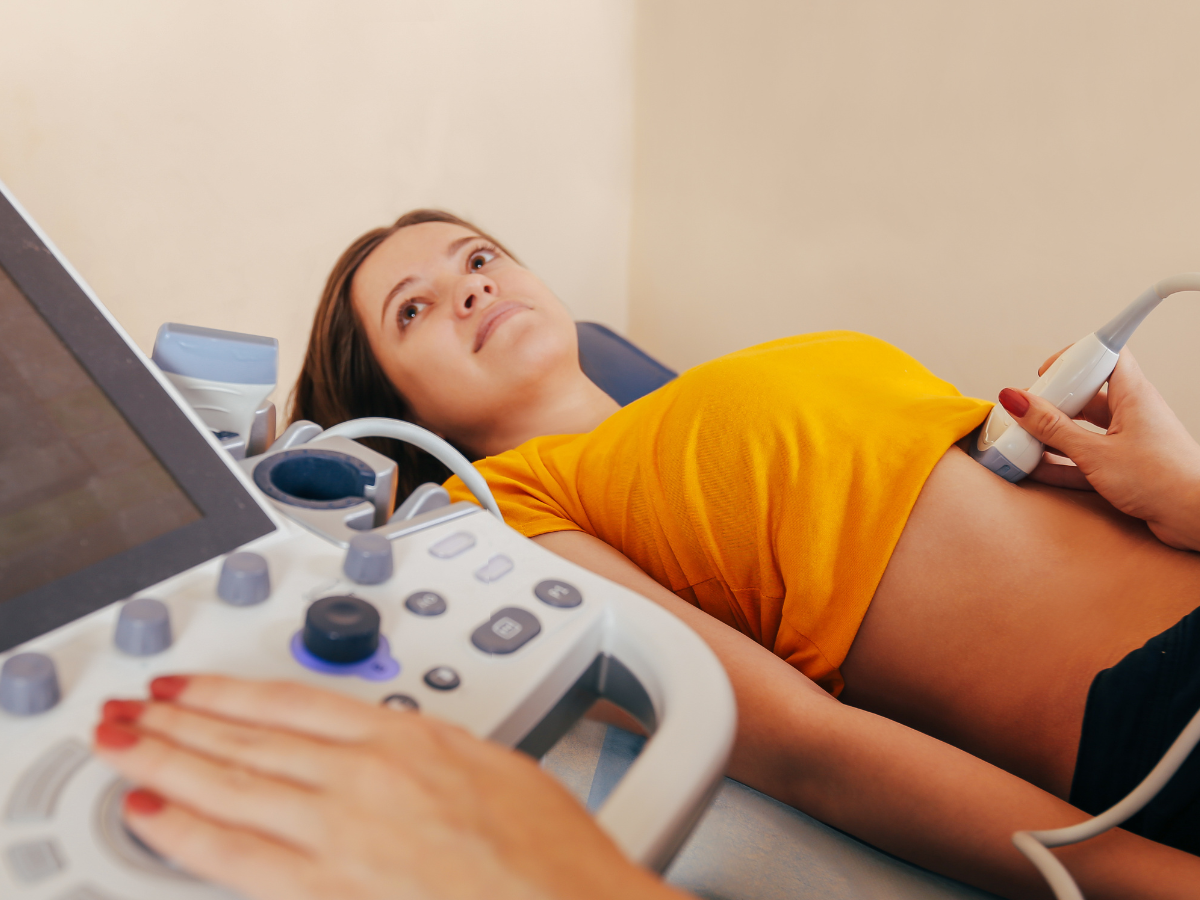
The Prostate SWE procedure is typically performed transrectally, similar to a standard transrectal ultrasound. While the procedure may cause some discomfort, it is generally well-tolerated and considered less invasive than other diagnostic methods like biopsy.
While Prostate SWE can provide valuable information about the stiffness of prostate tissue and help guide biopsy decisions, it does not replace the need for a biopsy in confirming the presence of prostate cancer. Biopsy remains the definitive method for diagnosing prostate cancer.
The procedure usually takes about 15 to 20 minutes to complete, depending on the specifics of the case. It is a relatively quick process that provides immediate results for analysis.
Typically, no special preparation is required for Prostate SWE. However, patients may be advised to follow specific instructions related to bowel preparation or to arrive with a full bladder, depending on the protocol of the imaging center.
Prostate SWE is a safe and non-invasive procedure with minimal risks. The most common side effects are temporary discomfort or a feeling of pressure during the transrectal ultrasound probe insertion. There is no exposure to ionizing radiation.
Prostate SWE has shown promise in increasing the accuracy of prostate cancer detection, especially in conjunction with traditional imaging and biopsy results. However, its effectiveness can vary based on the operator's experience and the specific characteristics of the prostate tissue being examined.