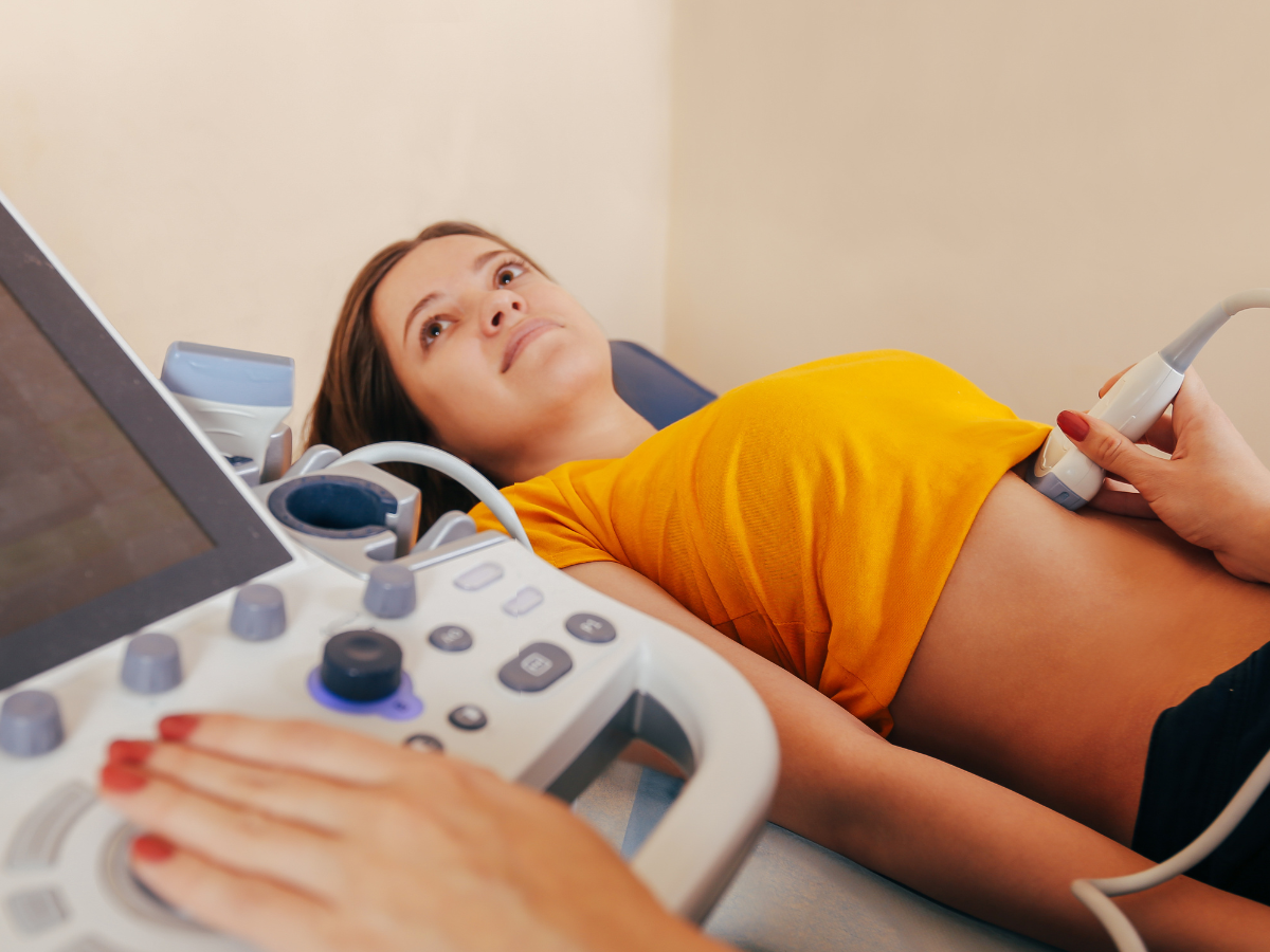
Prostate & Bladder Ultrasound Scan

Price : £215
Book an appoinment
A prostate and bladder ultrasound scan is a non-invasive diagnostic test used to assess the health and function of the prostate gland and bladder. It provides valuable insights into urological conditions such as benign prostatic hyperplasia (BPH), bladder abnormalities, and post-void residual urine volume. Importantly, this scan also aids in identifying suspicious changes that may indicate prostate or bladder cancer, making it a key tool in the early detection and management of urological malignancies.
✅ Related Ultrasound Scans:
What is a prostate ultrasound scan?
This scan combines the evaluation of the prostate gland and bladder to assess:
- 🔹 Prostate Size and Structure: Helps identify conditions like benign prostatic hyperplasia (BPH) or inflammation.
- 🔹 Bladder Health: Examines the bladder for abnormalities such as thickened walls, stones, or tumours.
- 🔹 Post-Void Residual Volume (PVR): Measures how much urine remains in the bladder after urination, which can indicate issues like urinary retention or obstruction.
The scan can be performed transabdominally (through the lower abdomen) or transrectally (for a more detailed prostate evaluation).
✅ Same-Day Appointments & Results 🚫 No GP Referral Needed 💳 Flexible Payment Options
Why is a Prostate & Bladder Ultrasound Important?
This ultrasound is critical for diagnosing and monitoring:
- 🔹 Benign Prostatic Hyperplasia (BPH): An enlarged prostate that can cause urinary difficulties.
- 🔹 Bladder Stones: Hard mineral deposits that can form in the bladder.
- 🔹 Urinary Retention: Difficulty emptying the bladder fully, often linked to prostate issues.
- 🔹 Bladder Tumours or Abnormalities: Including thickened bladder walls.
- 🔹 Prostate Health Concerns: Such as inflammation or other structural issues.
Who Should Consider a Prostate & Bladder Ultrasound?
This scan is recommended for men experiencing:
- 🔹 Urinary Symptoms: Difficulty starting or stopping urination, frequent urination, or weak urine flow.
- 🔹 Bladder Pain or Discomfort: Persistent pain or pressure in the bladder area.
- 🔹 Incomplete Bladder Emptying: Feeling like you can’t fully empty your bladder after urinating.
- 🔹 Enlarged Prostate Symptoms: Symptoms related to BPH or a family history of prostate conditions.
- 🔹 Routine Screening: Men over 50 or those with risk factors for prostate or bladder issues.
Benefits of a Prostate & Bladder Ultrasound Scan
- ✔ Early Detection of Prostate Issues:
- Detects prostate enlargement (benign prostatic hyperplasia, BPH), common in men as they age.
- Identifies tumours or masses in the prostate, aiding in the early detection of prostate cancer.
- Helps evaluate the prostate’s size and shape, providing clues about possible abnormalities.
- ✔ Assessment of Bladder Health:
- Detects bladder stones, which can cause pain or urinary problems.
- Identifies bladder wall thickening or irregularities that could indicate infections, inflammation, or cancer.
- Assesses how well the bladder empties after urination, identifying conditions like urinary retention.
- ✔ Monitoring and Management of Symptoms:
- Useful for men experiencing urinary problems, such as frequent urination, difficulty starting or stopping, or a weak stream.
- Helps diagnose obstructive conditions, like a blockage caused by an enlarged prostate.
🩺 Book Your Prostate & Bladder Ultrasound Scan Today
Take charge of your urological health with a prostate ultrasound scan. Early detection and management of prostate or bladder conditions can prevent complications and ensure long-term well-being. Schedule your scan today for expert care and peace of mind.
📞 Call us: 020 3318 1373
📧 Email: Contact@phoenix-ultrasound.co.uk
📍 Our Locations:
Central London: 1-5 Portpool Lane, London, Medical Room, EC1N 7UU
Surrey: 63 Nork Way, Banstead, SM7 1HL
FAQs
When I need a prostate and bladder ultrasound scan?
look for causes of persistent bladder problems - Urinary Incontinence - Blood in the urine - Bladder issues - Lower urinary tract infection -Urine that smells bad or is cloudy - Cystitis - Urinary retention -Increased urinary frequency especially at night - Feeling that your bladder has not emptied fully - Frequent urination - Strong, persistent urge to urinate - Burning sensation or pain when urinating - Signs of prostatism or enlarged prostate gland
What are signs of an enlarged prostate(Men)?
finding it difficult to start peeing. -straining to pee. -having a weak flow of urine. -quot; stop- start & quot; peeing. -Needing to pee urgently and/or frequently. -Needingng to get up frequently in the night to pee. -accidentally leaking urine (urinary incontinence)
How my private ultrasound scan can accelerate proceeding my case in NHS ?
Following the private ultrasound scan in phoenix ultrasound clinic, you can expect to receive your results within 24 hours. Subsequently, you can efficiently share these results with your General Practitioner (GP) or NHS healthcare team. This streamlined process facilitates the continuation of ongoing treatments or the initiation of new ones.
When will I get the results?
You will receive your results verbally immediately after the scan. Following the appointment, the sonographer will review the images and prepare a written report within 24 hours, with any recommended actions. The report and images will be sent to you, allowing you to discuss the findings with your doctor or physiotherapist if necessary.