Follicle count
Count the small follicles in each ovary.

A private pelvic ultrasound for polycystic ovaries (PCOS/PMOS) — follicle count, ovary size and the womb lining, explained clearly.
Ovaries, counted carefully.Choose your clinic and view appointment options in our online booking system.
Explore your appointmentHear from people
who have visited us.

PCOS is diagnosed from symptoms, blood tests and ultrasound together. The scan counts the small follicles in each ovary and measures the ovaries and lining.
Count the small follicles in each ovary.
Measure each ovary — enlarged ovaries are one PCOS feature.
Check the thickness of the lining, which can build up with irregular periods.
Look for cysts, fibroids or other causes of your symptoms.
A Polycystic Ovary Syndrome (PCOS) Ultrasound Scan is an essential diagnostic tool used to assess the presence of ovarian cysts and evaluate the overall health of the ovaries and reproductive system in women with suspected or diagnosed PCOS. PCOS is a common hormonal disorder that can affect fertility, menstrual cycles, and overall well-being. This non-invasive scan helps confirm the diagnosis of PCOS and guides treatment decisions.
Hear the summary of the text below from the following voice:
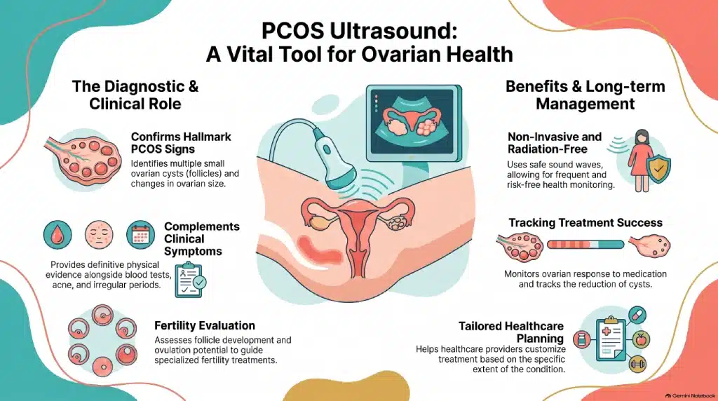
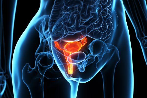
A PCOS Ultrasound Scan uses high-frequency sound waves to create detailed images of the ovaries and uterus. The scan looks for key signs of PCOS, such as the presence of multiple small ovarian cysts (follicles) and changes in ovarian size. These findings help confirm the diagnosis of PCOS, which is characterized by hormonal imbalances, irregular periods, and the potential presence of excess androgens (male hormones).
A PCOS Ultrasound plays a critical role in both the diagnosis and management of PCOS:
| Aspect | Details |
|---|---|
| Purpose | Assess the ovaries and uterus for signs associated with PCOS |
| Imaging Method | Ultrasound using high-frequency sound waves |
| Radiation | No radiation involved |
| What It Examines | Ovarian size, appearance, follicles, and uterine structures |
| Key Findings | Multiple small follicles and changes in ovarian morphology |
| Diagnostic Role | Supports PCOS assessment alongside symptoms, clinical history, and blood tests |
| Fertility Assessment | Evaluates follicle development and ovarian function |
| Treatment Monitoring | Helps monitor ovarian changes and response to treatment |
| Procedure | Non-invasive and generally well tolerated |
| Who May Benefit | Women with suspected or diagnosed PCOS, menstrual irregularities, or fertility concerns |
A PCOS Ultrasound Scan is recommended for individuals who:
If you suspect you have PCOS or need ongoing monitoring to manage your condition, a PCOS Ultrasound Scan provides essential information to guide your healthcare plan. Book your appointment today and take a proactive step towards managing your reproductive health.
From getting ready to discussing what comes next, here is a simple guide to your appointment.
A question before you book? Call usHow regular your periods are, symptoms such as acne or hair growth, and any blood test results.
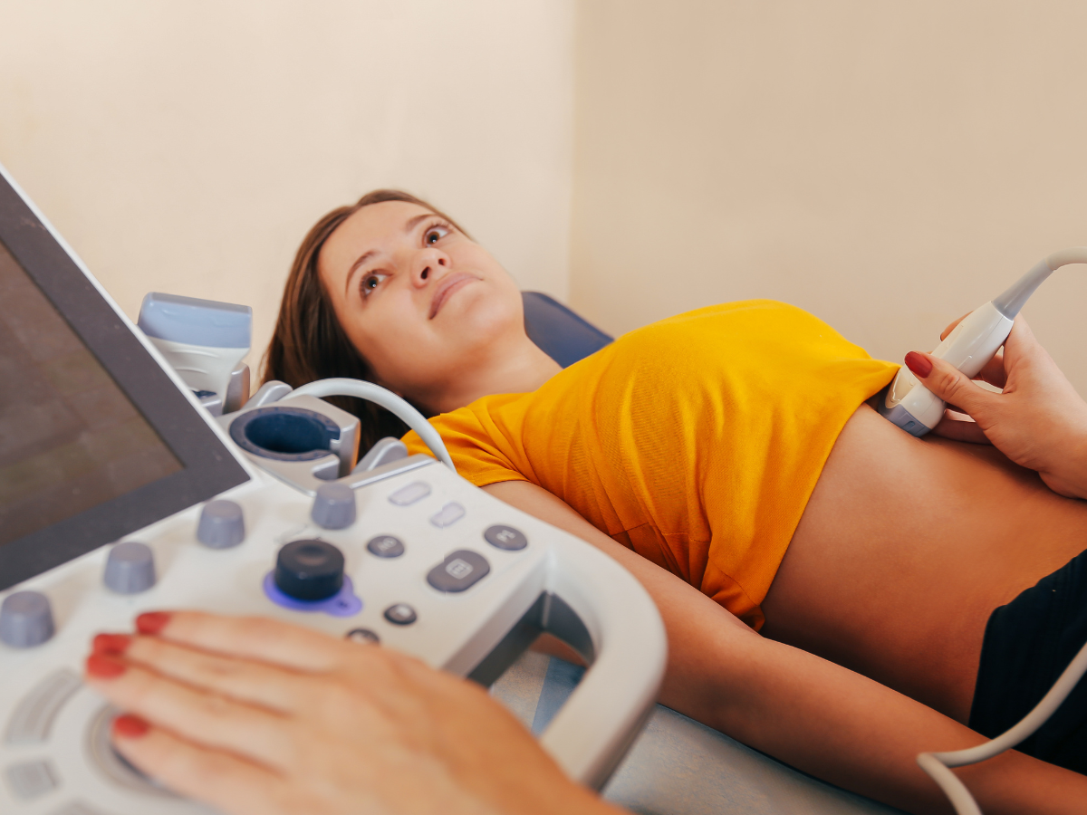
We usually use a slim internal probe for the clearest view, or scan over your tummy if you prefer. It is painless and takes about 20 minutes.
The sonographer talks you through what they have seen, and your written report follows within 24 hours — ready to share with your GP or specialist.
Choose the location that works for you. Confirm your selected clinic in the booking system before completing your appointment.
Need something more personal?
Speak to the team before booking.
No. You can book directly. If your GP or specialist has asked for this scan, bring their letter so the sonographer knows what to focus on.
No. Ultrasound is one of three criteria. Your doctor combines it with your symptoms and hormone tests.
Yes. We scan over your tummy instead, with a full bladder.
Yes. Where shown, the booking form offers a Full-body or Full-body Plus option that includes this area alongside the other organs, in one longer appointment.
A PCOS ultrasound scan is a medical imaging procedure used to diagnose polycystic ovary syndrome (PCOS) by visualizing the ovaries and identifying any cysts or other characteristic features.
It is performed to diagnose PCOS and to assess the size of the ovaries, the number of follicles, and the presence of cysts, which are common features of PCOS.
While an ultrasound scan can provide important information suggestive of PCOS, it is not the only diagnostic criteria. Your healthcare provider will also consider your medical history, symptoms, and possibly blood tests to make a diagnosis.
Yes, in addition to PCOS, ultrasound scans can detect other gynecological conditions such as ovarian cysts, fibroids, and endometriosis.
You will have a detailed report and images in 24 hours after your scan.
Use the booking form on this page and follow its Next buttons. Choose your scan option and appointment time, enter the requested details, then review the price before completing the booking.
Book your PCOS ultrasound below. Follow the booking form’s Next buttons to continue, one step at a time.
Loading the booking form…
Calendar not displaying? You can open the booking page in a new tab ↗.
Call the teamBooking this alongside another scan? Each scan keeps its own preparation. If anything is unclear, call us on 020 3318 1373 before your visit.
If you feel unwell, seek medical advice. This page is general information, not a diagnosis. For urgent concerns, contact your GP or NHS 111; in an emergency call 999.
Further reading: NHS guidance on PCOS
A PCOS ultrasound scan is a medical imaging procedure used to examine the ovaries and uterus. It can identify multiple small follicles and other ovarian features associated with polycystic ovary syndrome (PCOS).
A PCOS ultrasound scan is performed to assess the ovaries, identify features associated with PCOS, evaluate ovarian health, and support diagnosis and fertility assessment.
An ultrasound can show ovarian features commonly associated with PCOS, but it cannot diagnose PCOS on its own. Your healthcare provider will consider your symptoms, medical history, and blood test results alongside the ultrasound findings.
Please select Appointment type and Branch location to see time slots for booking- alternatively, please contact us via the chat, call or email, 020-3318-1373
Please check what preparation is needed before your ultrasound scan below:
| All Pelvic scans Kidney and Urinary tract Prostate & Urinary Tract Appendicitis Early Pregnancy Scan | Drink One Liter of still water (2 Pints) One hour before your scan. Do NOT use the toilet so you would arrive with a full bladder. Medication can be taken as normal. |
| Liver scans Upper Abdomen Liver Fibroscan (Liver Elastography) | Do NOT eat for 6 hours prior to scan time. |
| Combination of above scans | Same as respective scan’s preparation. |