Deep veins
Check the deep veins from groin to calf for clots.

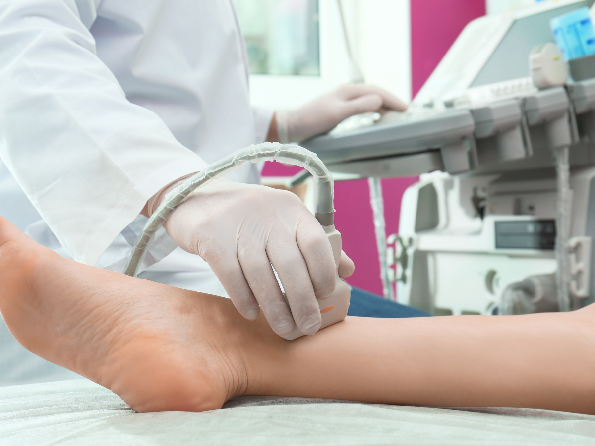
A private venous Doppler ultrasound to check for deep vein thrombosis (DVT) in the legs or arms — quick, painless and without radiation.
Veins, checked for clots.Choose your clinic and view appointment options in our online booking system.
Explore your appointmentHear from people
who have visited us.

A DVT is a blood clot in a deep vein, usually in the leg. Ultrasound checks whether each vein squeezes flat and whether blood flows normally.
Check the deep veins from groin to calf for clots.
Look for clots in surface veins (thrombophlebitis).
Assess veins after a previous DVT.
Baker’s cysts, muscle tears or swelling that can mimic a clot.
A Venous Doppler (DVT) ultrasound scan is a safe, non-invasive imaging test used to assess blood flow and detect blood clots in the deep veins of the legs or arms. It is commonly used to investigate leg swelling, calf or thigh pain, redness or warmth in a limb, and unexplained tenderness, particularly following surgery, prolonged immobility, or in those with a history of DVT or clotting disorders. The scan evaluates the deep and superficial veins, checks for the presence, location, and size of any clot, and assesses vein wall and valve condition. Our experienced sonographers provide clear, accurate results in a comfortable and private setting, with same-day appointments and results available and no GP referral needed.
If you are experiencing symptoms of DVT or have risk factors for venous thromboembolism, a Venous Doppler/DVT Ultrasound Scan is an essential step in protecting your health. Book your scan today with our experienced team to ensure timely diagnosis and treatment.
From getting ready to discussing what comes next, here is a simple guide to your appointment.
A question before you book? Call usSwelling, pain, recent travel, surgery, pregnancy or the pill.
Gel is applied and the probe is pressed gently along the vein. You may feel some pressure. It is painless and takes about 20 minutes.
The sonographer talks you through what they have seen, and your written report follows within 24 hours — ready to share with your GP or specialist.
Choose the location that works for you. Confirm your selected clinic in the booking system before completing your appointment.
Need something more personal?
Speak to the team before booking.
No. You can book directly. If your GP or specialist has asked for this scan, bring their letter so the sonographer knows what to focus on.
Choose one, two or three limbs in the booking form.
Sudden breathlessness, chest pain or coughing up blood can mean a clot has travelled to the lungs. Call 999. A painful, swollen leg should be checked the same day — if you cannot get a scan quickly, contact NHS 111.
Read NHS guidance ↗Symptoms of DVT: If you experience symptoms such as pain, swelling, and tenderness in one of your limbs, especially in the legs, a DVT Ultrasound Scan may be recommended to investigate the possibility of blood clot formation.
Medical Emergency: DVT is considered a medical emergency due to the potential complications it poses. Blood clots can lead to serious conditions, including pulmonary embolism if they travel to the lungs.
Risk Factors: Individuals with risk factors for DVT, such as a history of clotting disorders, recent surgery, prolonged immobility, or a family history of DVT, may undergo this scan as a preventive measure or to monitor their vascular health.
Post-Surgical Assessment: After certain surgeries, especially those involving the lower limbs, a DVT Ultrasound Scan may be performed to check for the development of blood clots.
Monitoring Treatment: For individuals already diagnosed with DVT, regular scans may be conducted to monitor the effectiveness of treatment and ensure the absence of new clot formation.
Unexplained Swelling or Pain: If you experience unexplained swelling or pain in your limbs, the scan helps identify the underlying cause, which could be DVT.
History of DVT: Individuals with a history of DVT may undergo periodic ultrasound scans to assess their ongoing risk and overall vascular health.
The most common symptoms of DVT include Pain, Swelling, and Tenderness in one of the limbs, usually in the legs.
DVT is a medical emergency because it can lead to serious complications, including the risk of blood clot movement to the lungs (pulmonary embolism), which can be life-threatening.
You will receive your results verbally immediately after the scan. Following the appointment, the sonographer will review the images and prepare a written report within 24 hours, with any recommended actions. The report and images will be sent to you, allowing you to discuss the findings with your doctor or physiotherapist if necessary.
Use the booking form on this page and follow its Next buttons. Choose your scan option and appointment time, enter the requested details, then review the price before completing the booking.
Book your DVT Doppler scan below. Follow the booking form’s Next buttons to continue, one step at a time.
Loading the booking form…
Calendar not displaying? You can open the booking page in a new tab ↗.
Call the teamBooking this alongside another scan? Each scan keeps its own preparation. If anything is unclear, call us on 020 3318 1373 before your visit.
If you feel unwell, seek medical advice. This page is general information, not a diagnosis. For urgent concerns, contact your GP or NHS 111; in an emergency call 999.
Further reading: NHS guidance on DVT
Please choose your appointment type, location, and time slot to book. If you need any help with booking, please contact us. We’re available 7 days a week, from 7am to 10pm.
Please check what preparation is needed before your ultrasound scan below:
| All Pelvic scans Kidney and Urinary tract Prostate & Urinary Tract Appendicitis Early Pregnancy Scan | Drink One Liter of still water (2 Pints) One hour before your scan. Do NOT use the toilet so you would arrive with a full bladder. Medication can be taken as normal. |
| Liver scans Upper Abdomen Liver Fibroscan (Liver Elastography) | Do NOT eat for 6 hours prior to scan time. |
| Combination of above scans | Same as respective scan’s preparation. |