Gap width
Measure the separation above, at and below the belly button.

A private diastasis recti ultrasound — measure the gap between your tummy muscles at rest and while you engage your core, and check for hernias.
Core muscles, measured.Choose your clinic and view appointment options in our online booking system.
Explore your appointmentHear from people
who have visited us.

After pregnancy or weight change, the tummy muscles can separate. Ultrasound measures the gap at several levels so you and your physiotherapist can track progress.
Measure the separation above, at and below the belly button.
Compare measurements when relaxed and when you lift your head.
Check the strength and thickness of the connective tissue.
Look for umbilical or abdominal-wall hernias.
A Diastasis Recti Ultrasound Scan is a specialised imaging test designed to assess the separation of the rectus abdominis muscles, commonly referred to as diastasis recti. This non-invasive procedure is crucial for diagnosing and evaluating the severity of this condition, particularly in postpartum women, but also in men and individuals following significant weight changes or abdominal surgeries. The scan helps determine the extent of the muscle separation and guides treatment planning.
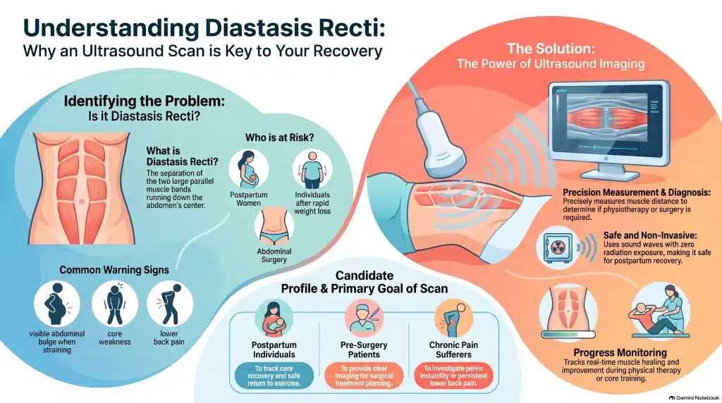
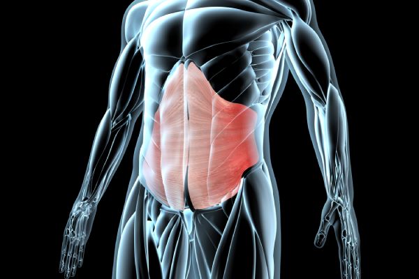
Diastasis recti is the separation of the two large parallel bands of muscles that run down the centre of the abdomen (rectus abdominis). This condition often occurs during pregnancy but can also result from excessive abdominal pressure in other situations.
Common symptoms include:
This scan plays a critical role in the diagnosis and management of abdominal muscle separation:
This scan is ideal for individuals who:
If you suspect you have diastasis recti or want to monitor your healing progress, this scan provides the insight and clarity you need. Book your scan today and receive expert care from our experienced sonographers.
From getting ready to discussing what comes next, here is a simple guide to your appointment.
A question before you book? Call usPregnancies, surgery, symptoms and what your physio has advised.
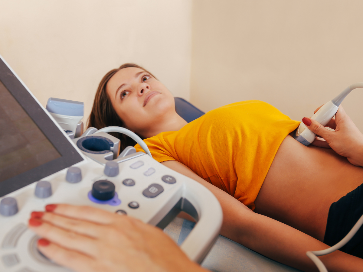
You lie on your back while gel is applied. We take measurements relaxed and while you gently lift your head. It is painless and takes about 20 minutes.
The sonographer talks you through what they have seen, and your written report follows within 24 hours — ready to share with your GP or specialist.
Choose the location that works for you. Confirm your selected clinic in the booking system before completing your appointment.
Need something more personal?
Speak to the team before booking.
No. You can book directly. If your GP or specialist has asked for this scan, bring their letter so the sonographer knows what to focus on.
Usually from 6–8 weeks after giving birth, or any time after if symptoms continue.
Yes. The booking form offers options with groin/hernia and upper abdominal and pelvic scans.
Use the booking form on this page and follow its Next buttons. Choose your scan option and appointment time, enter the requested details, then review the price before completing the booking.
Book your diastasis recti ultrasound below. Follow the booking form’s Next buttons to continue, one step at a time.
Loading the booking form…
Calendar not displaying? You can open the booking page in a new tab ↗.
Call the teamBooking this alongside another scan? Each scan keeps its own preparation. If anything is unclear, call us on 020 3318 1373 before your visit.
If you feel unwell, seek medical advice. This page is general information, not a diagnosis. For urgent concerns, contact your GP or NHS 111; in an emergency call 999.
Further reading: NHS guidance on hernias · NHS guidance on ultrasound scans
Please check what preparation is needed before your ultrasound scan below:
| All Pelvic scans Kidney and Urinary tract Prostate & Urinary Tract Appendicitis Early Pregnancy Scan | Drink One Liter of still water (2 Pints) One hour before your scan. Do NOT use the toilet so you would arrive with a full bladder. Medication can be taken as normal. |
| Liver scans Upper Abdomen Liver Fibroscan (Liver Elastography) | Do NOT eat for 6 hours prior to scan time. |
| Combination of above scans | Same as respective scan’s preparation. |