Liver and Gallbladder Ultrasound Scan

Private Liver and Gallbladder Ultrasound Scan
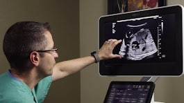
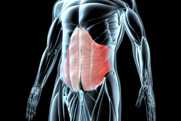
A liver and gallbladder ultrasound scan is a safe, non-invasive imaging procedure that provides detailed insights into the structure and function of both organs. It is widely used to assess abnormal liver function tests (e.g., elevated AST, ALT) and investigate symptoms like upper abdominal pain, jaundice, or digestive discomfort.
This scan helps detect a wide range of liver conditions, including fatty liver disease, cirrhosis, hepatitis, cysts, tumours, and alcohol-related damage. It also evaluates common gallbladder issues, such as gallstones, inflammation (cholecystitis), bile duct obstruction, and polyps. Early detection of liver and gallbladder diseases can significantly improve treatment outcomes.
🔍 What is a Liver and Gallbladder Ultrasound Scan?
This ultrasound uses high-frequency sound waves to produce real-time images of your liver, gallbladder, and surrounding structures. It evaluates:
- ✔ Fatty Liver Disease (Hepatic Steatosis): Detects excess fat in the liver tissue.
- ✔ Liver Cirrhosis: Identifies scarring and fibrosis from long-term liver damage.
- ✔ Liver Tumours and Cysts: Detects benign or malignant growths within the liver.
- ✔ Inflammation and Infections: Checks for hepatitis, liver abscess, or other infections.
- ✔ Gallstones (Cholelithiasis): Identifies stones within the gallbladder or bile ducts.
- ✔ Cholecystitis: Detects inflammation of the gallbladder wall.
- ✔ Gallbladder Polyps: Finds small growths inside the gallbladder lining.
- ✔ Bile Duct Obstruction: Evaluates blockages that may cause jaundice or infection.
📌 Why is it Important?
- ✔ Fatty Liver Detection: Especially important for patients with obesity, diabetes, or metabolic syndrome.
- ✔ Liver Damage Assessment: Evaluates alcohol-induced or viral damage (e.g., hepatitis).
- ✔ Tumour & Cancer Screening: Enables early detection and diagnosis of liver or gallbladder abnormalities.
- ✔ Gallstone Diagnosis: Identifies the cause of upper abdominal pain, especially after meals.
- ✔ Pre-Surgical or Pre-Treatment Evaluation: Helps with planning for surgeries or liver-related interventions.
👤 Who Should Consider This Scan?
- ✔ Persistent pain in the upper right abdomen
- ✔ Symptoms of gallstones (pain after fatty meals, nausea, vomiting)
- ✔ Jaundice, dark urine, or pale stools
- ✔ Unexplained weight loss or fatigue
- ✔ History of liver disease, hepatitis, or gallbladder problems
- ✔ Abnormal liver function tests (LFTs) or suspected gallbladder pathology
💡 Benefits of a Liver and Gallbladder Ultrasound
- ✔ Non-invasive, safe, and radiation-free
- ✔ Early detection of liver and gallbladder diseases
- ✔ Supports diagnosis of abdominal pain and digestive symptoms
- ✔ Same-day appointments with fast, accurate results
🩺 Book Your Liver and Gallbladder Ultrasound Today
Whether you’re experiencing symptoms or simply want peace of mind, our private ultrasound scans provide expert evaluation with immediate results. Available in Central London and Surrey (Banstead).
📞 Call us: 020 3318 1373
📧 Email: Contact@phoenix-ultrasound.co.uk
📍 Our Locations:
Central London: 1-5 Portpool Lane, London, Medical Room, EC1N 7UU
Surrey: 63 Nork Way, Banstead, SM7 1HL
📊 Add-On Option: Liver Shear Wave Elastography (FibroScan)
You can also book Liver Shear Wave Elastography (also known as FibroScan), a quick, painless test that measures liver stiffness and fat content. It helps detect early stages of fibrosis or steatosis and is ideal for ongoing monitoring of chronic liver conditions.


