A pelvic ultrasound scan is a safe, radiation-free imaging test that checks your uterus, ovaries and pelvic cavity, usually completed in 15–30 minutes, with a verbal summary on the day and no GP referral required.
Hear the summary of the text below from the following voice:
| Your situation | Best scan for you | Quick rule |
|---|---|---|
| Heavy or irregular periods, pelvic pain, abnormal bleeding | Diagnostic pelvic ultrasound (transabdominal and/or transvaginal) | No GP referral needed — book directly |
| Routine gynaecological health check, no symptoms | Well Woman Pelvic Scan | Best for general screening and peace of mind |
| IVF preparation or cycle tracking | Fertility Pelvic Scan | Assesses follicles and uterine baseline anatomy |
| Any of the above | Private scan from £145–£220 | Same or next-day appointments available |
Definition
A pelvic ultrasound scan is a non-invasive, radiation-free diagnostic imaging test that lets a sonographer assess your uterus, endometrium (womb lining), ovaries, fallopian tubes and the wider pelvic cavity in real time. Because it uses sound waves rather than X-rays, it is one of the safest ways to investigate gynaecological symptoms or check pelvic wellbeing at any age.
This guide covers the female pelvic ultrasound scan. If you are looking for the male equivalent, see our abdominal and pelvic ultrasound for men service, or read our blog on what a male pelvic ultrasound shows.
When and Why We Use Pelvic Ultrasound
Pelvic ultrasound serves two broad purposes: investigating symptoms, and checking wellbeing or fertility even when nothing feels wrong.
Common Symptoms and Clinical Indications
Patients typically come to us for irregular or heavy periods, abnormal bleeding (including after menopause), pelvic pain, bloating, pain during intercourse, or fertility investigations. We also use pelvic ultrasound to monitor known conditions such as fibroids, ovarian cysts, PCOS, pelvic inflammatory disease (PID), endometriosis, or to confirm correct IUD placement.
| Symptom | May indicate | Typical next step |
|---|---|---|
| Heavy or irregular periods | Fibroids, adenomyosis, hormonal imbalance | Pelvic scan, bloods if advised by your GP |
| Abnormal or post-menopausal bleeding | Endometrial thickening, polyps | Prompt scan and GP discussion |
| Pelvic pain or bloating | Ovarian cysts, PCOS, PID | Pelvic scan, Doppler if indicated |
| Painful intercourse | Endometriosis, fibroids | Pelvic scan, specialist referral if needed |
| Fertility concerns | Follicle count, uterine anatomy | Fertility Pelvic Scan |
If you have a red-flag symptom such as post-menopausal bleeding, or your GP suspects a gynaecological cancer, please see your GP before booking a private scan. This ensures you’re also placed on the correct urgent NHS referral pathway alongside any private imaging you choose to have.
Transabdominal vs Transvaginal Scan
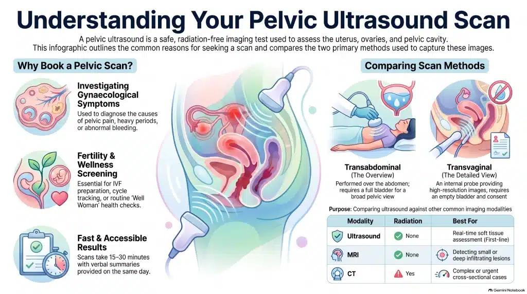
Two methods are used, often together. Transabdominal scanning is performed over a full bladder for a broad overview of the pelvis. Transvaginal scanning uses an internal probe for sharper detail and always requires your consent and an empty bladder. Your sonographer will recommend the most appropriate method and adjust based on your comfort at every step.
| Transabdominal | Transvaginal | |
|---|---|---|
| Preparation | Full bladder (drink ~1 litre beforehand) | Empty bladder |
| Image detail | Broader overview | Higher detail, especially ovaries and endometrium |
| Consent | Standard | Explicit consent required |
| Best for | Initial overview, patients preferring a non-internal scan | Detailed assessment, fertility work-up |
Screening and Fertility Context
Not every pelvic scan is prompted by symptoms. A Well Woman Pelvic Scan checks general anatomy and screens for anomalies as part of routine health maintenance. A Fertility Pelvic Scan instead maps baseline uterine anatomy and follicle count or development — useful ahead of IVF or when tracking a natural cycle.
| Well Woman Pelvic Scan | Fertility Pelvic Scan | |
|---|---|---|
| Focus | General anatomy and anomaly check | Follicle count and uterine baseline |
| Best timing | Any point in your cycle | Specific cycle day, as advised |
| Suits | Routine wellbeing check | IVF preparation, cycle tracking |
Benefits vs Other Imaging Modalities
Ultrasound’s main advantages are speed, zero radiation, and strong accuracy for assessing the uterus and ovaries. Its limitations are that very small lesions, and deep infiltrating endometriosis in particular, can be harder to fully characterise by ultrasound alone — MRI is often used alongside or instead for a complete picture in these cases. Where blood flow matters — for example with a cyst or fibroid — your sonographer may add Doppler imaging, and 3D imaging is available when clinically useful.
| Modality | Radiation | Typical time | Best for |
|---|---|---|---|
| Ultrasound | None | 15–30 minutes | Real-time soft tissue assessment, first-line |
| MRI | None | 30–60 minutes | Small or deep lesion detail |
| CT | Yes | 10–20 minutes | Complex or urgent cross-sectional cases |
What Your Scan Includes
Every appointment begins with a consultation with a qualified sonographer, who discusses your history and explains the process before you choose, with consent, between transabdominal and transvaginal scanning, or both. The scan itself assesses your uterus, endometrial lining, ovaries, adnexa and pelvic cavity, with Doppler or 3D imaging added where clinically appropriate. Most appointments last 15–30 minutes. You receive a verbal summary immediately, followed by a secure digital report and images via patient portal or email. If you are currently under the care of another clinician, we recommend discussing whether this scan is necessary with them first.
What to Expect on the Day
Wear a two-piece outfit so your abdomen is easy to access. For a transabdominal scan, drink around one litre of water an hour beforehand and avoid emptying your bladder; for a transvaginal scan, empty your bladder first. A water-based gel is used throughout, and you may feel mild pressure, though the scan itself should not be painful. Your sonographer will talk you through each step and share findings as they go.
Who Performs the Scan & Equipment Used
All scans are performed by HCPC-registered sonographers or consultant radiologists, using modern duplex or 3D/4D ultrasound systems with Doppler capability. Many of our specialists also work within the NHS, bringing extensive women’s health imaging experience to every private appointment.
Safety, Limitations & Contraindications
Pelvic ultrasound has no known side effects and carries none of the risks associated with ionising imaging. According to the NHS:
“Ultrasound scans do not use any radiation, unlike in X-rays or CT scans.”
Image quality can occasionally be reduced by bowel gas or higher body weight, and there are very few true contraindications beyond ensuring comfort and informed consent for any internal scan. If a scan is inconclusive, we may recommend further imaging such as MRI.
Pricing & Booking
Private pelvic ultrasound scans in London typically range from £145 to £220, depending on clinic, scan type, and any add-ons such as Doppler or 3D imaging. No GP referral is required for routine self-referral, and same-day or next-day appointments are usually available online or by phone (see the note above on when to see your GP first). We accept a range of payment methods and insurance, and our cancellation and refund policy is available at the time of booking. Combined Well Woman or Fertility packages are also available for a more comprehensive assessment.
| Booking type | What it covers | Typically falls |
|---|---|---|
| Single scan (TA or TV only) | One method, standard assessment | Lower end of the £145–£220 range |
| Combined TA + TV | Both methods for full detail | Mid-range |
| With Doppler or 3D add-on | Extra imaging where clinically useful | Upper end |
| Well Woman / Fertility package | Comprehensive assessment, extra checks | Upper end |
Locations
We see patients at two locations: our central London clinic in Holborn, and our Banstead clinic in Surrey — both offering the same scan quality, equipment, and same or next-day availability.
References & Governance
Our pelvic ultrasound protocols follow guidance from the NHS and the British Medical Ultrasound Society (BMUS), the UK’s professional body for ultrasound practice. The clinic is CQC-registered and handles all patient data in line with UK GDPR. Every scan goes through internal quality assurance, and any findings that need further input trigger a prompt referral to a gynaecologist or GP.
Our Clinicians & Specialist Team
Our senior sonographers and consultant radiologists hold HCPC registration and NHS affiliations, bringing years of women’s health imaging experience to every appointment. Where a scan reveals a finding that needs further assessment, we trigger an immediate referral pathway to a gynaecologist or your GP, so nothing is left waiting.
Consultation & Aftercare
You can discuss your results with the sonographer immediately after your scan and ask any questions on the spot. Depending on the findings, we recommend the appropriate next steps, whether that’s reassurance, a follow-up scan, or referral for treatment.
Leave a Reply