A private breast ultrasound in London images both breasts and the axillary lymph nodes using sound waves alone — no radiation — with a verbal summary on the day and a full written report following within 12–24 hours.
Hear the summary of the text below from the following voice:
| Your situation | Recommended scan | Quick rule |
|---|---|---|
| New lump, pain, or nipple discharge | Bilateral Breast & Axilla Ultrasound | First-line test under 40; same-day appointments available |
| Family history or general peace of mind | Bilateral Breast & Axilla Ultrasound | Not a replacement for NHS screening, but fills the gap between rounds |
| Known cyst or lump needing more detail | Breast Ultrasound + Shearwave Elastography | Measures tissue stiffness to help judge if follow-up is needed |
| Breast implants (suspected rupture) | Bilateral Ultrasound, +3D option available | Ultrasound is first-line for implants — no compression involved |
| Pregnant or breastfeeding with a symptom | Standard Breast & Axilla Ultrasound | Safe at any stage — uses no ionising radiation |
Definition
A breast ultrasound scan (also called breast USG) uses high-frequency sound waves — not X-rays — to build a real-time picture of the breast tissue, the nipple-areolar area, and the lymph nodes in the axilla (armpit). A probe is moved over gel-covered skin, and the returning echoes are turned into an image the sonographer reads immediately.
The scan does two main jobs: it characterises abnormalities — telling a solid mass from a fluid-filled cyst — and it checks the axillary lymph nodes whenever a symptom or diagnosis needs staging. Because it uses sound waves rather than radiation, it’s a safe, repeatable test, including for women with dense breast tissue, where mammography alone can miss changes.
Mammography and MRI remain valuable, but each modality answers a different question: ultrasound is strongest at telling cystic from solid, MRI at whole-breast surveillance in very high-risk cases, and mammography at picking up the micro-calcifications ultrasound can’t see. The comparison below sets out how the three fit together.
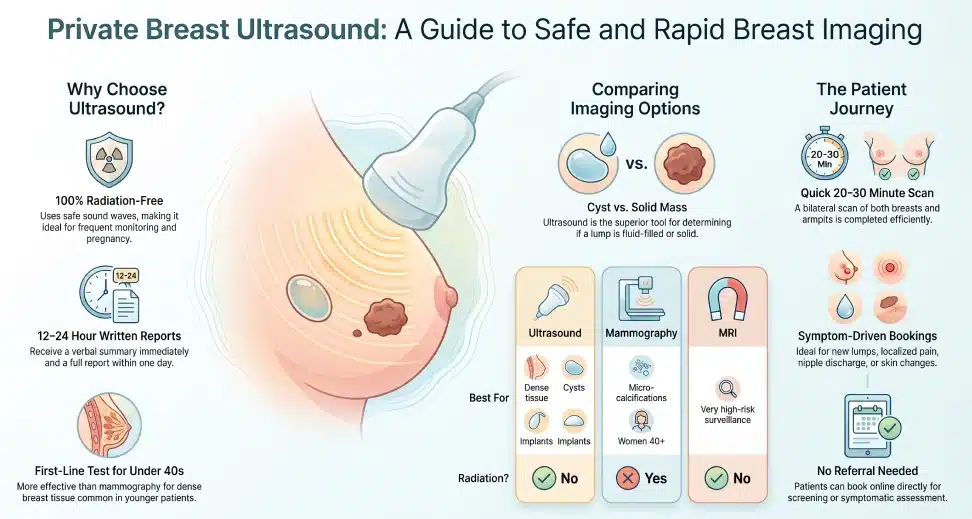
When and Why We Use Breast Ultrasound
Breast ultrasound is rarely used on its own — it answers a specific clinical question, whether that’s investigating a symptom, screening a higher-risk patient, or resolving something a mammogram couldn’t settle.
Clinical Indications & Symptoms
Most private bookings are symptom-driven. Common reasons to book quickly include a new lump or thickening, nipple discharge, localised pain, a change in skin texture or dimpling, or an abnormal finding picked up on examination or a previous scan. Features such as a rapidly enlarging lump, new nipple inversion, or a new lump in the armpit warrant prompt assessment, which is why same-day private appointments exist. Ultrasound is the first-line test under 40; from 40 onwards, a new lump is normally assessed with mammography and ultrasound together (sometimes called triple assessment alongside clinical examination), since ultrasound alone can miss the micro-calcifications mammography is better at catching.
Screening / Peace of Mind Use
Not every booking follows a symptom. Women with a family history of breast cancer, those feeling anxious between routine screening rounds, or anyone who simply wants a baseline check can book a bilateral scan for reassurance. This doesn’t replace the NHS Breast Screening Programme, but it fills the gap for people outside the invited age range, or those who want more frequent monitoring.
Complement to Mammography & MRI
| Modality | Uses radiation? | Best for | Limitation |
|---|---|---|---|
| Ultrasound | No | Dense breasts, cyst vs solid, implants, pregnancy/breastfeeding | Can miss micro-calcifications |
| Mammography | Yes (low dose) | Micro-calcifications, women 40+, national screening | Less accurate in dense tissue |
| MRI | No | Very high-risk surveillance, problem-solving | Higher cost, longer waits, less available |
Ultrasound and mammography are complementary rather than competing tests — a finding on one is often clarified by the other, and neither replaces the fuller picture your GP or breast specialist builds over time.
Special Populations
Three groups benefit particularly from ultrasound-first imaging: women under 35, whose denser tissue limits mammography; pregnant or breastfeeding women, for whom avoiding radiation matters; and women with breast implants, where ultrasound is the recommended first step for suspected rupture or capsular contracture, without the compression a mammogram applies.
What Your Scan / Service Includes
A bilateral breast and axilla scan examines all four quadrants of both breasts plus both underarms, so left and right can be directly compared — useful, since asymmetry is itself a diagnostic clue.
| Imaging plane | What it shows |
|---|---|
| Radial | Follows the natural duct pattern — useful for nipple discharge |
| Anti-radial | Cross-checks the radial view from a different angle |
| Transverse | Side-to-side view for overall tissue mapping |
| Longitudinal | Top-to-bottom view, often paired with transverse for a lesion’s full extent |
The sonographer records each plane, grades any lesion using standard descriptors — margins, vascularity, internal echoes, solid versus cystic — and takes measurements for the report. Colour Doppler is added when blood flow through a lesion needs assessing. You get a verbal summary immediately, then a full written report, typically within 12–24 hours, with images you can share with your GP or specialist. Comparing against a previous mammogram, or arranging biopsy guidance, can usually be added to the same visit.
What to Expect on the Day
Wear a two-piece outfit — it’s far easier to access the chest and underarm than in a one-piece dress. After registering and giving verbal consent, you’ll change into a gown and lie on the couch, usually on your back with the arm on the side being scanned raised above your head. The sonographer applies a warm gel and moves the probe over the breast and axilla; you may feel light pressure, but it shouldn’t hurt — say so if it does. A bilateral scan takes around 20–30 minutes. Afterwards you wipe off the gel, get dressed, and the sonographer talks you through what they saw before you leave, with the full report to follow.
Who Performs the Scan & Equipment Used
Scans are performed by HCPC-registered sonographers and consultant-level radiologists with a specific interest in breast imaging. Equipment includes high-frequency linear probes and colour Doppler as standard, plus — for cases needing finer characterisation — GE ABUS (Automated Breast Ultrasound) and shear wave elastography, which measures tissue stiffness as an extra clue to whether a lesion needs following up. All machines undergo regular maintenance, calibration, and quality-assurance checks.
Safety, Limitations & Contraindications
Breast ultrasound has an excellent safety record because it uses no ionising radiation at all — a genuine advantage over mammography for repeated or frequent scanning. According to Cancer Research UK:
“It doesn’t involve radiation and there are usually no side effects.”
That said, it isn’t the right tool for every question: ultrasound can miss the micro-calcifications that sometimes flag very early cancer, and image quality can be reduced by obesity or overlying bowel gas in some scan positions. If a finding is suspicious or inconclusive, you’ll usually be advised to have a mammogram, a biopsy, or both. Beyond an open wound or active skin infection over the area to be scanned, there are essentially no contraindications, and adverse effects are extremely rare.
Pricing & Booking
| Scan option | Price |
|---|---|
| Breast ultrasound (one side) | £215 |
| Breast & axilla ultrasound (both sides) | £265 |
| Breast & axilla ultrasound + shearwave elastography (both sides) | £355 |
| 3D breast & axilla ultrasound (both sides) | £355 |
| 3D breast & axilla ultrasound + shearwave elastography (both sides) | £445 |
Unilateral scans suit a single, one-sided symptom; bilateral is the more common choice for comparison, screening, or family-history monitoring, and the axillary lymph nodes are included as standard on every option. No GP referral is needed — book online or by phone, with same-day appointments available for symptomatic patients. Flexible payment, including interest-free instalments, is available, and rescheduling follows the clinic’s standard cancellation policy.
Locations
| Branch | Address | Nearest transport |
|---|---|---|
| Central London (Holborn) | 1–5 Portpool Lane, London, EC1N 7UU | Chancery Lane and Farringdon tube stations are both about a 4-minute walk; Bus 55 and 243 stop on Rosebery Avenue. Note: this branch does not currently have step-free access. |
| Banstead, Surrey | 63 Nork Way, Banstead, SM7 1HL | Bus 164, a 7-minute walk from Banstead Station; on-site parking available |
Both branches take bookings by phone or online, with phone and text support available seven days a week, 07:00–22:00.
References & Governance
This page draws on guidance from the NHS, Cancer Research UK, and the Royal College of Radiologists. The clinic operates under Care Quality Commission (CQC) registration, and all sonographers and radiologists are individually registered with the Health and Care Professions Council (HCPC). Patient data is handled under UK GDPR.
Our Clinicians & Specialist Team
Scans are read by consultant radiologists and HCPC-registered sonographers who specialise in breast and axillary imaging, with well over a decade of ultrasound experience across NHS and private practice.
[Note for Amin/client: no named clinician bios were publicly available to source accurately — insert real names, subspecialty, years of experience, and any BMUS/RCR/EFSUMB memberships here once supplied, to strengthen this section’s E-E-A-T.]
Aftercare, Follow-Up & Escalation Pathways
| What the scan shows | Typical next step |
|---|---|
| Normal / no abnormality | No further action; return if a new symptom appears |
| Fluid-filled cyst | Usually just monitored; if drainage is appropriate, you’ll be referred for a fine-needle aspiration |
| Solid lump, low suspicion | Mammogram comparison and/or interval follow-up scan |
| Solid lump, higher suspicion | Biopsy referral, arranged promptly |
If you’d like to discuss your results or next steps in more detail, you can book a follow-up consultation with the reporting specialist directly, without going through your GP first.
Leave a Reply