Prostate Transrectal Colour Doppler Ultrasound

Price : £365
Book an appoinment
Prostate Transrectal Colour Doppler Scan
A Prostate Transrectal Colour Doppler Scan is a specialised ultrasound procedure designed to assess the health and condition of the prostate gland. Using advanced Doppler imaging technology, this scan provides detailed information about blood flow and potential abnormalities within the prostate, helping with the early detection of prostate issues, such as enlargement, inflammation, or prostate cancer.
What is a Prostate Transrectal Colour Doppler Scan?
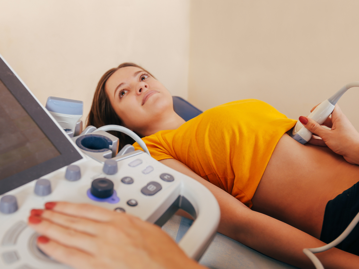
This scan is a type of transrectal ultrasound (TRUS) that uses a small probe inserted into the rectum to produce high-resolution images of the prostate. The colour Doppler feature measures the blood flow within and around the prostate, providing crucial information on:
- Prostate size and shape
- Blood flow patterns (which can help identify abnormal growths)
- Presence of prostate lesions or tumours
- Vascularisation (how much blood supply is reaching certain areas)
✅ Related Ultrasound Scans:
Why is a Prostate Transrectal Colour Doppler Scan Important?
This scan is especially useful for diagnosing and monitoring conditions such as:
- Benign Prostatic Hyperplasia (BPH): An enlarged prostate that can cause urinary problems.
- Prostatitis: Inflammation or infection of the prostate.
- Prostate Cancer: The scan helps in early detection by analysing abnormal blood flow patterns.
- Post-Treatment Monitoring: After prostate surgery or treatment, this scan can help monitor recovery and ensure there are no residual issues.
Who Should Consider a Prostate Transrectal Colour Doppler Scan?
This scan is recommended for men who:
- Experience symptoms such as frequent urination, difficulty starting or stopping urination, or weak urine flow.
- Have elevated PSA (Prostate-Specific Antigen) levels, which can indicate prostate issues.
- Are undergoing prostate cancer screening or need ongoing monitoring.
- Have a family history of prostate problems and want proactive screening.
Benefits of Prostate Transrectal Colour Doppler Scan
- Non-invasive: Unlike biopsies, this scan offers a non-invasive way to detect prostate issues.
- High Accuracy: The colour Doppler function provides a precise assessment of blood flow and potential abnormalities.
- Early Detection: Helps identify prostate issues at an early stage, which is crucial for successful treatment.
Book Your Prostate Transrectal Colour Doppler Scan Today
Early detection is key to effective prostate health management. Book your prostate transrectal colour Doppler scan with us to ensure timely diagnosis and personalised care. Our experienced specialists will guide you through every step, ensuring your comfort and safety.
📞 Call us: 020 3318 1373
📧 Email: Contact@phoenix-ultrasound.co.uk
📍 Our Locations:
Central London: 1-5 Portpool Lane, London, Medical Room, EC1N 7UU
Surrey: 63 Nork Way, Banstead, SM7 1HL
FAQs
What is the purpose of a Prostate Transrectal Colour Doppler Ultrasound, and when is it recommended?
A Prostate Transrectal Colour Doppler Ultrasound is an advanced imaging procedure designed to evaluate the prostate gland's , size, structural changes and blood flow , aiding in the diagnosis and monitoring of various prostate conditions, including cancer. It is recommended based on specific clinical indications and your healthcare provider's assessment.
How can I schedule a Prostate Transrectal Colour Doppler Ultrasound at Phoenix Ultrasound Clinic?
To arrange a Prostate Transrectal Colour Doppler Ultrasound, please book the scan online or contact our appointment hotline at 08000485738. Our knowledgeable staff will guide you through the scheduling process and assist in securing a suitable time for your examination.
Is it necessary to book an appointment, or can I walk in for the Prostate Transrectal Colour Doppler Ultrasound?
While we strive to accommodate walk-ins when possible, scheduling an appointment ensures a more organized and personalized experience. We recommend contacting our clinic to arrange a convenient time for your procedure.
What information is required when booking an appointment for a Prostate Transrectal Colour Doppler Ultrasound?
When scheduling your appointment, please provide essential personal details, including your name, contact information, and any relevant medical history related to prostate health. This information helps customize the examination to your specific needs.
What does the Prostate Transrectal Colour Doppler Ultrasound procedure involve?
During the procedure, a specialized ultrasound probe is gently inserted into the rectum to obtain detailed images of the prostate. The use of Colour Doppler technology allows for the assessment of blood flow within the prostate gland
How long does a Prostate Transrectal Colour Doppler Ultrasound typically take?
The average procedure taking approximately 20 minutes. The complexity of the examination may influence the overall time required.
Is any special preparation required before a Prostate Transrectal Colour Doppler Ultrasound?
Generally, you may be instructed to attend with full bladder to get transabdominal scans of your pelvis and then empty your bladder before the transrectal scan .
How are the results of the Prostate Transrectal Colour Doppler Ultrasound communicated, and what is the expected timeframe?
The results will be meticulously interpreted by our qualified ultrasound specialist. You will receive a comprehensive report.
In case of any concerns or questions regarding my Prostate Transrectal Colour Doppler Ultrasound, what should I do?
If you have any concerns or questions, please don't hesitate to contact our dedicated hotline at 08000485738 .Our customer service team is committed to addressing your inquiries and ensuring a professional and reassuring experience at Phoenix Ultrasound Clinic.
How my private ultrasound scan can accelerate proceeding my case in NHS ?
Following the private ultrasound scan in phoenix ultrasound clinic, you can expect to receive your results within 24 hours. Subsequently, you can efficiently share these results with your General Practitioner (GP) or NHS healthcare team. This streamlined process facilitates the continuation of ongoing treatments or the initiation of new ones.
When will I get the results?
You will receive your results verbally immediately after the scan. Following the appointment, the sonographer will review the images and prepare a written report within 24 hours, with any recommended actions. The report and images will be sent to you, allowing you to discuss the findings with your doctor or physiotherapist if necessary.