A DVT ultrasound scan (venous Doppler) is a painless, radiation-free test that checks the deep leg veins for a blood clot, usually with same-day results.
If you have sudden breathlessness, chest pain or are coughing blood alongside leg swelling, call 999 or go to A&E now. These can signal a pulmonary embolism and need emergency assessment — not a booked private scan.
Hear the summary of the text below from the following voice:
| Your question | The short answer | Quick rule |
|---|---|---|
| What does it check for? | Blood clots in the deep leg veins (DVT), their location and extent | No clot found = vein flows and compresses normally |
| Does it hurt or use radiation? | No — it’s a painless, radiation-free ultrasound | Uses sound waves only, safe to repeat |
| How long does it take? | 20–45 minutes depending on scope | One leg is quicker than both legs or a full-leg scan |
| When should I book urgently? | Same-day if you have new leg swelling, pain or redness | Call 999 if you also have chest pain or breathlessness |
What Is a DVT Ultrasound Scan?
Deep vein thrombosis (DVT) is a blood clot in one of the deep veins, most often in the calf or thigh. A duplex (Doppler) ultrasound scan is the standard, non-invasive way to examine these veins, combining B-mode imaging with colour Doppler to show whether blood is flowing normally or a clot is blocking the vessel. Its job is threefold: detect a clot, map its location and extent, and confirm the vein still compresses and carries blood normally. Because it uses sound waves rather than X-rays, the test is safe and can be repeated as often as needed.
When and Why We Use Ultrasound Scan for DVT
Ultrasound is requested whenever DVT is suspected clinically, whether symptoms are mild and nagging or sudden and alarming. Below we cover the warning signs, how UK practice triages suspected cases, and how duplex scanning compares with the alternatives.
Symptoms & Risk Triggers
DVT symptoms typically affect one leg only — bilateral swelling is more often something else. Risk rises after recent surgery, long immobility (including long-haul flights), pregnancy, obesity, a previous DVT, or an inherited clotting tendency.
| Sign or risk factor | What it may indicate |
|---|---|
| Swelling, pain or tenderness in one calf | Classic DVT presentation — book a scan promptly |
| Skin redness, warmth or discolouration | Inflammation over an affected vein segment |
| Sudden breathlessness or chest pain | Possible pulmonary embolism — treat as a medical emergency |
| Recent surgery, flight or bed rest | Temporary immobility risk factor |
| Pregnancy, obesity or prior DVT | Longer-term or recurring risk factor |
Care Pathway & Triage (NICE-style)
UK practice uses the Wells score to estimate how likely DVT is before any scan is booked, combined with a D-dimer blood test where appropriate. Higher-risk patients are prioritised for an urgent scan. According to NICE, people with a likely DVT Wells score should:
“have a proximal leg vein ultrasound scan within 4 hours of it being requested”
Private same-day scanning is designed for suspected DVT without emergency features — it runs alongside this NHS pathway, not instead of it. Anyone with red-flag symptoms should go straight to A&E rather than wait for a booked appointment.
Benefits vs Alternatives
Duplex ultrasound is first-line because it’s quick, non-invasive and repeatable. Its limits: severe swelling or very muscular legs can make some vein segments harder to visualise.
| Test | Radiation | Speed | Best for |
|---|---|---|---|
| Duplex/Doppler ultrasound | None | Same-day, 20–45 min | First-line test for almost all suspected DVT |
| CT venography | Yes | Same-day, hospital setting | Inconclusive ultrasound or pelvic/abdominal extension |
| MR venography | None | Longer, specialist referral | Complex or recurrent cases needing detailed mapping |
What Your Scan/Service Includes
Every scan is tailored to what’s requested — a single leg, both legs, or a full assessment to the calf veins.
| Element | Details |
|---|---|
| Scope | Common femoral, femoral and popliteal veins; full-leg assessment to the calf veins if indicated; one or both legs |
| Technique | B-mode imaging, vein compressibility testing, colour and pulse-wave Doppler for flow assessment |
| Output | Same-day verbal explanation, plus a full digital report and high-resolution images sent securely |
| Duration | 20–45 minutes depending on unilateral, bilateral or whole-leg scope |
What to Expect on the Day
Registration is quick. Wear comfortable clothing allowing easy access to the leg. The sonographer applies gel and uses gentle pressure along the vein to test compressibility — a healthy vein flattens easily, a clotted one resists. You may shift position slightly for the best imaging angle, then get an immediate summary of findings, with the full written report following shortly after.
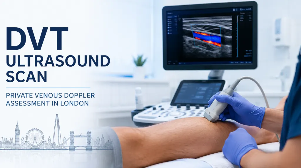
Who Performs the Scan & Equipment Used
Scans are carried out by HCPC-registered sonographers or consultant radiologists, using advanced duplex and Doppler systems for high-resolution vein imaging. Several clinicians also work within the NHS.
Safety, Limitations & Contraindications
The scan is safe, painless and radiation-free, so it can be repeated without cumulative risk. Image quality can be reduced by severe swelling, a limb cast, or open wounds over the area examined. If a clot extends into the pelvis, or findings are inconclusive, further imaging such as CT or MR venography may be recommended.
Pricing & Booking
Most London clinics price a private DVT ultrasound within a similar bracket; bilateral scans cost more since more vein territory is assessed.
| Scan type | Typical London price range |
|---|---|
| Unilateral (one leg) | Roughly £150–£250 |
| Bilateral (both legs) | Roughly £250–£450 |
Booking is available online or by phone, with same-day or next-day appointments in most cases. We accept major payment cards; some private medical insurers also cover this scan when referred by a GP. Please check our cancellation policy when booking, as short-notice changes may incur a fee.
Locations
| Location | Address | Contact | Phone & text support | Nearest transport |
|---|---|---|---|---|
| Central London (Holborn) | 1-5 Portpool Lane, London, Medical Room, EC1N 7UU | 020 3318 1373 | Mon–Sun, 07:00–22:00 | Chancery Lane (5-min walk); Farringdon (10-min walk); bus 55/243 from Rosebery Avenue |
| Banstead, Surrey | 63 Nork Way, Banstead, SM7 1HL | 020 3318 1373 | Mon–Sun, 07:00–22:00 | Bus 164, 7-min walk from Banstead Station; on-site parking |
References & Governance
Duplex and Doppler ultrasound for suspected DVT follows established UK clinical standards, including compressibility protocols and first-line imaging guidance in NICE’s guideline on venous thromboembolic diseases (NG158). Clinics of this kind are regulated by the CQC, with scans performed by HCPC-registered practitioners.
Our Clinicians & Specialist Team
Our sonographers and consultant radiologists are HCPC-registered, and several also hold NHS posts. If a scan confirms a DVT, we make sure you understand the next step — same-day referral for anticoagulation, or follow-up with a vascular specialist — so nothing is left unclear.
Leave a Reply