A soft tissue lumps and bumps scan is a same-day ultrasound that shows, within minutes, whether a new lump is a harmless cyst or something that needs further checking — with no needles, no radiation and usually no GP referral required.
Hear the summary of the text below from the following voice:
| Your question | Quick answer |
|---|---|
| What is it? | An ultrasound scan of a lump and the surrounding skin, fat, muscle, tendon, nerves and vessels |
| Do I need a referral? | No — self-referral is available; a GP referral is also accepted |
| How long does it take? | Usually 15–30 minutes |
| What does it cost? | From around £130 for a single area |
| When do I get results? | Verbal summary same day; written report and images follow by email |
Definition
A soft tissue lumps and bumps scan is a diagnostic ultrasound examination of the superficial tissues of the body — skin, fat, muscle, connective tissue, blood vessels and nerves — used to assess a palpable lump, swelling or bump. It’s non-invasive and uses high-frequency sound waves to build a real-time image, so there’s no ionising radiation involved at any point. The scan gives an immediate picture of the lump’s size, shape and internal structure, which is the starting point for working out what it is and what, if anything, needs to happen next.
When and Why We Use Soft Tissue Lumps & Bumps Scan
Most people come to us because they’ve noticed a lump, swelling or bump somewhere on the body and want to know what it is. A soft tissue scan is appropriate whether the lump is brand new, has been there a while but is changing, or has appeared after an injury. You can be referred by your GP, or you can book directly yourself in a private setting.
| Presentation | What we look at | Why it matters |
|---|---|---|
| New lump | Size, shape, solid vs fluid-filled | Rules out concerning features early and often provides reassurance |
| Growing or changing lump | Change over time, blood flow (Doppler) | Growth or increased vascularity can indicate a need for further imaging |
| Painful bump | Tenderness, inflammation, fluid content | Helps identify a cause such as a cyst, abscess or haematoma |
| Recurrent bump | Recurrence pattern, scarring from before | Guides longer-term management or the case for removal |
| Post-trauma swelling | Bleeding, fluid collection | Confirms a haematoma and its size versus other soft tissue injury |
Whatever the trigger, the aim is the same: characterise the lump — is it solid or cystic? — and use that to guide next steps, whether that’s simple reassurance and monitoring, a referral to a specialist, or further imaging.
What the Scan Includes
The scan covers the lump itself plus the surrounding soft tissue layers, so any nearby vessels, nerves or tendons that could be relevant are assessed too. Preparation is minimal for almost everyone — you don’t need to fast or change your routine, just wear clothing that allows easy access to the area.
| Scan type | Typical duration | Preparation |
|---|---|---|
| Single lump or bump | 15–20 minutes | None required |
| Multiple lumps or a wider area | 20–30 minutes | None required |
| Abdominal soft tissue (uncommon) | 20–30 minutes | Fasting occasionally advised — you’ll be told in advance |
You’ll leave with still images from the scan, and in many cases digital access to those images alongside your written report, along with a clear steer on any recommended next steps.
What to Expect on the Day
You’ll check in, and the sonographer will take a brief history and note the specific clinical question your scan needs to answer. A small amount of gel is applied to the skin and the probe is moved gently over the area of concern — you may be asked to change position or expose the area a little further so the whole lump can be seen clearly. The procedure itself is painless, though you might notice some light pressure, particularly if the lump is already tender.
Where appropriate, the sonographer can show you the live images and talk you through what they’re seeing as the scan progresses. At the end, you’ll typically get a verbal summary of the findings before you leave, with the full written report and images following by email or online access — some clinics can turn this around the same day. If the lump isn’t clearly seen, or the findings suggest further imaging would help, you’ll be advised on the best next step.
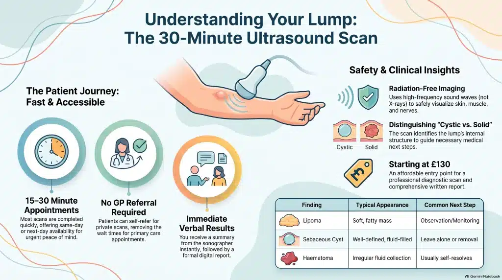
Who Performs the Scan & Equipment Used
Soft tissue scans are carried out by HCPC-registered sonographers or consultant radiologists with specific experience in musculoskeletal and soft tissue ultrasound. The equipment used is a high-resolution diagnostic ultrasound machine with a linear high-frequency transducer, which is what gives the sharp detail needed for superficial structures close to the skin. Where blood flow needs to be assessed — for instance, to check the vascularity of a solid mass — Doppler ultrasound is used alongside the standard greyscale images.
Conditions Detected / Diagnostic Value
The vast majority of lumps referred for scanning turn out to be entirely benign. Ultrasound is well suited to telling these common, harmless findings apart, and to flagging the minority of cases where a solid mass, unusual blood flow or rapid growth means something warrants closer attention.
| Common finding | Typical ultrasound appearance | Usual next step |
|---|---|---|
| Lipoma | Soft, well-defined fatty mass | Usually monitored; rapid growth, pain, or size over 5cm prompts MRI to rule out liposarcoma |
| Sebaceous cyst | Well-defined, sometimes with a central punctum | Can be left alone or removed if it recurs or becomes infected |
| Ganglion cyst | Fluid-filled, near a joint or tendon | Often observed, or aspirated if uncomfortable — though recurrence after aspiration is common |
| Haematoma | Irregular fluid collection, usually post-injury | Typically resolves on its own; re-scan if it isn’t settling |
| Solid mass with atypical features | Irregular margins, increased vascularity | Referral for MRI and/or biopsy to confirm what it is |
Beyond these, the scan can also pick up foreign bodies, post-traumatic swelling and, in some cases, hernias. What ultrasound does particularly well is answer the first, most useful question — cystic or solid? — which is exactly why it’s used as the first-line imaging test for the great majority of lumps and bumps.
Safety and Limitations
Ultrasound uses sound waves rather than ionising radiation, which makes it a genuinely safe, non-invasive way to look inside the body — it’s why it’s the default first step for almost any new lump. That said, it isn’t the right tool for every situation. Very deep lesions, or those sitting behind bone, may not be fully visualised. A higher BMI, overlying gas, or awkward anatomy can all reduce image clarity. Ultrasound is also operator-dependent, so the quality of the images and how they’re interpreted can vary with the sonographer’s experience and how complex the area is. Because of this, ultrasound cannot say with certainty on its own whether a lump is benign or malignant — some findings need MRI or biopsy to reach a firm answer, and results are always read alongside your symptoms and history by the referring clinician. A soft tissue scan is a diagnostic adjunct: it supports your GP or specialist’s assessment, rather than replacing it, and it isn’t a substitute for an urgent cancer-pathway referral (such as a 2-week-wait referral) where your GP feels one is needed.
Pricing & Booking
Private soft tissue lumps and bumps scans in London typically sit in the following price bands, though the exact figure depends on how many areas are examined and whether a specialist appointment is included.
| Option | Typical price | What’s included |
|---|---|---|
| Single-area scan | From around £130 | Ultrasound of one lump/bump, verbal summary, written report |
| Additional areas | Extra charge per area | Applies where more than one lump is examined in the same visit |
| Scan + specialist doctor appointment | Around £260 | Same-day scan plus consultant review and same-day results |
The written report is included in every price above — there’s no separate charge for it. Booking is available online or by phone, and self-referral is welcomed — you don’t need to see your GP first. Same-day or next-day appointments are often available for private scans.
Locations
Scans are available at our Central London clinic in Holborn, as well as our Banstead, Surrey location, both chosen for easy access whether you’re coming from central London or Surrey. Evening and weekend appointments can be arranged at certain times, and both sites are well served by public transport.
Our Clinicians & Specialist Team
Our sonographers and consultant radiologists bring experience from both NHS and private practice, with specific training and accreditation in musculoskeletal and soft tissue ultrasound. Every clinician is registered with the HCPC or the relevant professional body, and the team is committed to clear, patient-friendly communication throughout your appointment — so you leave understanding exactly what was found and what it means.
Patient Experience
Patients consistently tell us the same things matter most: a comfortable environment, minimal waiting, and being talked through the findings in plain language. Most people get a verbal summary on the day, along with digital or emailed access to their images and report shortly afterwards. If you’d like your results reviewed with your GP or a specialist, we can help arrange that too, with clear guidance on any recommended follow-up so nothing is left hanging.
References & Governance
Our soft tissue ultrasound service is regulated by the Care Quality Commission (CQC) and follows national ultrasound safety guidance from the British Medical Ultrasound Society (BMUS), keeping acoustic exposure as low as reasonably practicable — the safety principle that applies to ultrasound, as distinct from the ALARA principle used for X-ray and CT. All practitioners are registered with the Health & Care Professions Council (HCPC) or an equivalent body and follow their professional standards. According to RadiologyInfo.org, a patient resource maintained by the Radiological Society of North America and the American College of Radiology:
“Most soft-tissue masses are harmless, although some may be cancerous.”
This is exactly why a soft tissue scan is useful even for lumps that turn out to be nothing — it replaces uncertainty with a clear answer. The scan is intended to complement NHS care and your GP’s assessment, not to replace a necessary referral or treatment.
Leave a Reply