A neck ultrasound is a painless, radiation-free scan that uses high-frequency sound waves to examine the thyroid, lymph nodes, salivary glands and blood vessels in your neck, usually completed in under 30 minutes with same-day feedback.
Hear the summary of the text below from the following voice:
| What you want to know | Quick answer |
|---|---|
| What is it? | A soft-tissue ultrasound of the neck — no radiation, no needles |
| How long does it take? | 15–30 minutes |
| GP referral needed? | No — self-referral available |
| Price from | £130 |
| Results | Verbal feedback same day; written report within 24–48 hours |
| Preparation | None — just avoid high collars, necklaces and thick facial hair over the scan area |
Definition
A neck ultrasound — sometimes called an ultrasound of the neck, a neck scan ultrasound, or ultrasound on neck — is a non-invasive imaging scan that uses high-frequency sound waves to visualise the soft tissues of the neck, including the lymph nodes, salivary glands, thyroid gland and carotid arteries, and to identify any abnormalities in those structures. Because it doesn’t use ionising radiation, it can be repeated safely whenever your clinician needs an updated picture, and it’s often the first imaging test recommended for a neck lump, swelling or thyroid concern.
When and Why We Use Neck Ultrasound
We recommend a neck ultrasound whenever a lump, swelling or unexplained symptom points to the soft tissues of the neck.
| If you have… | We’re checking for… |
|---|---|
| A lump or swelling in the neck | Cysts, reactive nodes, tumours or salivary gland masses |
| Persistent hoarseness or difficulty swallowing | Thyroid enlargement, nodules or masses pressing on nearby structures |
| Abnormal thyroid blood tests | Structural thyroid disease — nodules, goitre, thyroiditis |
| Enlarged or unusual lymph nodes | Infection, inflammation or, rarely, malignancy |
| Suspected salivary gland problem | Stones, infection or tumours in the parotid or submandibular glands |
| Risk of carotid artery narrowing | Plaque, stenosis and flow changes linked to stroke risk |
We also use neck ultrasound to follow up known nodules or masses over time, tracking any change in size or appearance as part of ongoing clinical management.
What the Scan Includes
Depending on your referral, your scan may cover one or more of the following structures.
| Structure | What we look at |
|---|---|
| Lymph nodes | Size, shape and any reactive or suspicious change |
| Salivary glands (parotid, submandibular) | Stones, infection or tumours |
| Thyroid and parathyroid (if included) | Nodules, goitre, thyroiditis |
| Carotid arteries (vascular protocol) | Narrowing, plaque and blood flow |
| Superficial masses | Whether a lump is solid, cystic or vascular |
A full neck scan typically takes 15–30 minutes and needs no special preparation beyond removing necklaces, loosening a high collar, and trimming a beard if it covers the area being examined. Your images and report are usually delivered electronically the same day or within 24–48 hours. If you specifically need thyroid imaging, check whether this is included in your scan or booked as a separate thyroid-and-neck package — we’ll confirm this when you book.
What to Expect on the Day
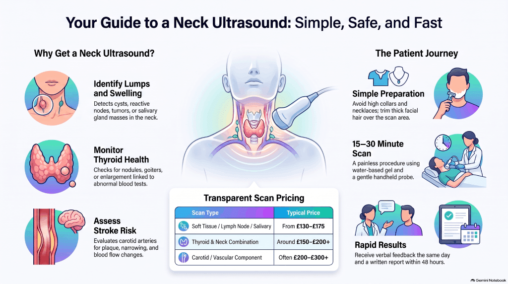
You’ll check in and may be asked to fill in a short history and consent form. You’ll then lie on the scan couch with your neck slightly extended while water-based gel is applied to the skin — necklaces and high collars need to come off, and if a beard covers the scanning area you may be asked to trim it. The sonographer moves the probe gently over your neck; you’ll feel mild pressure but no pain. Once the scan is finished you can return to normal activities immediately, with no downtime. Many patients get verbal feedback on the day, followed by a full written report by email within 24–48 hours; we’ll arrange a follow-up consultation if one is needed.
Who Performs the Scan & Equipment Used
Your scan is performed by a HCPC-registered diagnostic sonographer or a consultant radiologist with specific experience in head and neck, thyroid or vascular imaging. We use modern high-frequency linear probes (typically 7–15 MHz), with colour Doppler and duplex vascular imaging available for carotid assessments. Our clinic is CQC-inspected, and images are reviewed by a consultant radiologist where clinically required. Our team combines NHS experience with dedicated private-practice expertise in head and neck diagnostics.
Conditions Detected / Diagnostic Value
Neck ultrasound is highly sensitive for the soft tissues of the neck and is commonly used to identify:
| Area | Common findings |
|---|---|
| Thyroid | Nodules (solid or cystic), goitre, thyroiditis (Hashimoto’s/Graves’), features suspicious for thyroid cancer |
| Lymph nodes | Reactive, infective or, less commonly, metastatic/primary malignant change |
| Salivary glands | Stones, sialadenitis, parotid or submandibular tumours |
| Neck masses | Benign cysts, soft tissue masses, vascular malformations |
| Carotid arteries | Stenosis, plaque and flow abnormalities relevant to stroke risk |
We can also use ultrasound to guide a needle biopsy of a nodule or lymph node that needs further sampling. Ultrasound is excellent for superficial soft tissue, but some deeper or bony structures are better assessed with CT or MRI — we’ll advise if further imaging is needed.
Safety and Limitations
Neck ultrasound uses sound waves rather than ionising radiation, so it’s safe, non-invasive and well tolerated by almost everyone. According to Guy’s and St Thomas’ NHS Foundation Trust:
“There are no known risks to ultrasound imaging.”
That said, ultrasound has real limits worth understanding before you book.
| Ultrasound does well | Ultrasound is limited by |
|---|---|
| Soft tissue detail: nodes, glands, superficial masses | Bone and air-filled structures, which it can’t penetrate effectively |
| Real-time, radiation-free imaging | Very small lesions hidden deep or behind bone |
| Repeat scans with no cumulative risk | Body habitus — deeper structures can be harder to image in larger patients |
| Image-guided biopsy | Operator and equipment dependence — choose an accredited, experienced clinic |
If a finding is inconclusive, your sonographer or radiologist may recommend a CT or MRI scan to complete the picture.
Pricing & Booking
Pricing depends on which structures are included in your scan.
| Scan type | Typical price |
|---|---|
| Soft tissue / lymph node / salivary gland scan | From £130–£175 |
| Thyroid and neck combination scan | Around £150–£200+ |
| Carotid / vascular component added | Often £200–£300+ |
These ranges can overlap because they reflect different combinations of structures scanned — your final price depends on exactly what’s included, and we’ll confirm it before your appointment.
You can self-refer for a private neck ultrasound — a GP referral usually isn’t required — and same-day or next-day appointments are often available, including evening and weekend slots at some locations. Payment options, insurance acceptance and our cancellation policy are confirmed at the time of booking. Book online or call us to schedule your scan.
Locations
We see patients at our central London clinic in Holborn and at our Banstead clinic in Surrey, both chosen for convenient access by public transport with nearby parking. Both locations offer step-free access and a chaperone on request, with evening and weekend appointments available at some sites — let us know when you book which slots suit you best.
Our Clinicians & Specialist Team
Our lead radiologists and senior sonographers bring NHS backgrounds alongside dedicated head and neck imaging experience, and many hold membership of professional bodies such as the British Medical Ultrasound Society and the Royal College of Radiologists. We work closely with ENT and vascular specialists so that any abnormal finding has a clear next step. Our team brings together NHS-level expertise within a private environment focused on prompt diagnosis and patient comfort.
Patient Experience
We prioritise fast access — same-day or next-day appointments and quick turnaround reporting — in comfortable, private scan rooms where our team talks you through every step. You’ll receive your images and written report electronically, with the option of a GP or specialist referral if follow-up is needed. Our pricing is transparent with no hidden fees, and if a scan raises questions, we’ll guide you toward the right next step, whether that’s an ENT, endocrine or vascular specialist.
Leave a Reply