A PCOS ultrasound scan is a pelvic imaging test that checks the ovaries and uterus for the follicle count, ovarian volume and endometrial pattern used to help diagnose polycystic ovary syndrome.
Hear the summary of the text below from the following voice:
| Your question | Quick answer | Detail |
|---|---|---|
| Do I need a GP referral? | No | Self-refer online, by phone or by email |
| External or internal scan? | Your choice | Transvaginal (internal) is more detailed; transabdominal (external) needs a full bladder |
| How long does it take? | 15–20 minutes | Plus registration and a findings discussion |
| What does it cost? | From about £185–£200 | £690–£990 for a package with consultation and bloods |
| When are results ready? | Same day or next day | Written report and images sent electronically |
Definition
A PCOS (Polycystic Ovary Syndrome) ultrasound is a specialised scan focused on the ovaries and uterus, used to assess features commonly seen in PCOS such as multiple small follicles and increased ovarian volume. It can be performed as an abdominal ultrasound for PCOS (transabdominal, external) or as a transvaginal scan (internal), depending on clinical need and personal preference. Many patients search for it simply as PCOS scanning or a PCOS ultrasound scan, and the two terms describe the same test.
When and Why We Use PCOS Ultrasound
A PCOS ultrasound scan is typically referred or requested for one of a few recurring reasons, each pointing the sonographer toward a slightly different focus during the scan.
| Symptom or reason | What it may suggest | What the scan assesses |
|---|---|---|
| Irregular or absent periods | Possible anovulation | Follicle pattern and endometrial lining |
| Excess facial/body hair, acne, scalp thinning | Possible androgen excess | Ovarian volume and morphology |
| Fertility difficulty | Irregular or absent ovulation | Follicle development and ovulation signs |
| Known PCOS, ongoing care | Response to treatment | Change in follicle count or volume over time |
| Pelvic symptoms, unclear cause | Another gynaecological issue | Fibroids, endometriosis, cysts |
What the Scan Includes
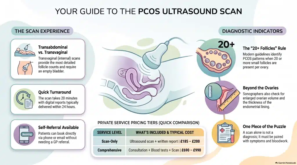
The scan examines both ovaries — follicle count and ovarian volume — along with the uterine lining and structure, and usually takes 15–20 minutes depending on scan type and clinical indication. A written report with images is generated and sent electronically or given on-site.
| Scan type | Preparation | Best for | What to expect |
|---|---|---|---|
| Transabdominal (external) | Full bladder | Initial screening, patient preference | Gel applied to the lower abdomen; non-invasive |
| Transvaginal (internal) | Bladder usually empty | A more detailed follicle count | Slender probe, typically very well tolerated; a female sonographer can be requested |
What to Expect on the Day
You’ll arrive, register, and briefly go through your medical history and symptoms with the sonographer. For an external scan, you lie on the couch while gel and the transducer are applied to your lower abdomen; for an internal scan (with your consent), a slender probe is used for more detailed imaging. Afterwards, the sonographer can usually talk you through the initial findings, and your full report — often same-day or within 24 hours — follows by email or in person. The environment is private and unhurried, and you’re encouraged to ask questions; results can then be reviewed with your GP, gynaecologist or fertility specialist.
Who Performs the Scan & Equipment Used
Scans are carried out by HCPC-registered sonographers or consultant radiologists specialising in women’s health imaging, using high-resolution ultrasound machines capable of both transabdominal and transvaginal probes for accurate follicle counts and ovarian measurements. The service is delivered under rigorous clinical governance and quality standards.
Conditions Detected / Diagnostic Value
The scan is built to identify the sonographic features of polycystic ovarian morphology, while also checking for other explanations of your symptoms.
| Feature | Typical finding | PCOS-associated pattern |
|---|---|---|
| Follicles per ovary | Fewer, varied sizes | 20 or more small follicles (2–9mm) per ovary on modern high-frequency transvaginal ultrasound, often around the periphery |
| Ovarian volume | Within normal range | Enlarged ovarian volume |
| Endometrial lining | Typical for cycle stage | Can appear thickened alongside irregular cycles |
| Other pelvic findings | — | May instead reveal fibroids, cysts or endometriosis |
This follicle-count threshold follows the 2023 international evidence-based PCOS guideline; the original 2003 Rotterdam criteria used a lower cut-off of 12 or more follicles, which some clinics using older or lower-resolution equipment still reference. The scan also monitors change over time — follicle development or response to ovulation induction — for patients already managing PCOS.
Safety and Limitations
Ultrasound uses sound waves rather than radiation, so it’s safe to repeat as often as clinically needed. A transvaginal scan is generally more accurate for follicle counting than a transabdominal one, and image quality can be affected by body habitus, bladder fullness and where you are in your menstrual cycle. Ultrasound also isn’t used to diagnose PCOS within the first eight years after a first period, since a multi-follicular ovary pattern is a normal stage of adolescent development rather than a sign of PCOS. Crucially, ovarian appearance is only one part of a PCOS diagnosis. According to the Royal College of Obstetricians and Gynaecologists:
“Having polycystic ovaries on an ultrasound does not necessarily mean that you have PCOS.”
That’s why a scan is read alongside your symptoms and, usually, blood tests — never as a stand-alone verdict.
Pricing & Booking
Private PCOS ultrasound pricing in London generally falls into two tiers, and most clinics offer same-day or next-day appointments without needing a GP letter first.
| Package | What’s included | Typical London price |
|---|---|---|
| Scan-only | PCOS ultrasound scan and written report | ~£185–£200 |
| Comprehensive package | Consultation, blood tests and scan | ~£690–£990, depending on clinic and scope |
Booking is usually available online, by telephone, or via email, with self-referral often possible. you can check price list for other services.
Locations
PCOS ultrasound scans are available across London, including central locations such as Harley Street and Welbeck Street, alongside satellite clinics in Greater London — most are easily reached by public transport, with parking or a nearby tube station close by.
Our Clinicians & Specialist Team
Your scan and results sit within a team of experienced consultant gynaecologists, fertility specialists and HCPC-registered sonographers, many with NHS backgrounds and specialist training in women’s reproductive imaging and PCOS management. Clear clinical governance means a straightforward referral pathway into endocrinology, fertility or gynaecology services if your results need follow-up.
Patient Experience
Compared with a standard NHS referral route, private booking means shorter waits and same-day or next-day reports, with digital access to your images and PDF report as standard. The setting is private and unhurried, with clear next-step guidance — whether that’s a referral, lifestyle advice or an ongoing monitoring plan — given before you leave.
References & Governance
This service reflects UK guidance that ultrasound is one component of a PCOS diagnosis, alongside clinical history and hormone testing — never the sole basis for it. Clinics operate under CQC regulation, maintain sonographer accreditation, and use up-to-date imaging equipment, with every scan reviewed by qualified clinicians and results communicated appropriately to your referring practitioner.
Leave a Reply